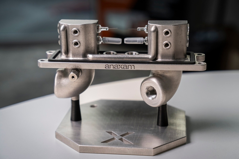

An environment and vacuum chamber was customized for the experiments to condition the pre-filled syringes for neutron imaging. Credit: Paul Scherrer Institute
In extremely uncommon cases, the needles of prefilled syringes might end up being obstructed. This can have possibly destructive effects for clients if their medication does not get in the body or the dose is too low.
A group from the Paul Scherrer Institute PSI, led by the ANAXAM innovation transfer center in partnership with MSS, has actually now had the ability to take a comprehensive appearance inside such needles. The scientists had the ability to identify the possible reasons for the clogs and develop the conditions to avoid them in the future.
The secret was a mix of imaging strategies at the Swiss Light Source SLS and the neutron source SINQ. Both lie in close distance to each other on the PSI website– an ensemble that is distinct worldwide. The outcome is an initially, state chemical engineer Vladimir Novak and physicist and CEO of ANAXAM Christian Grünzweig, “We have actually prospered in taking the most comprehensive appearance yet at the within a needle.”
Pre-filled syringes (PFS) are progressively utilized for cancer treatment, for instance, utilizing monoclonal antibodies. In a little number of cases, the needles of the pre-filled syringes block. The threat of this has actually increased considering that the restorative representatives, which include proteins and antibodies, have actually ended up being more focused and, for that reason, more thick, providing higher resistance to fluid circulation.
No dependable figures are readily available on the typical frequency of a clog. Scientists are talking about ever-changing pressure or temperature level conditions as possible causes, for instance, while transferring batches.
Pressure and temperature level variations as possible causes
The group led by chemical engineer Vladimir Novak, head of the job group and scientist at ANAXAM, examined this hypothesis in a series of measurements. 31 PFS were exposed to modifications in temperature level in between 5 and 40 degrees and modifications in pressure in between 550 and 1010 millibars. Such conditions generally take place throughout flights when transferring medical items. The group took a look at the needles utilizing both synchrotron X-ray and neutron beams.
Both imaging techniques have particular benefits. Neutrons can permeate metals much better however are deflected by hydrogen atoms, which happen in liquids and hence produce a clear contrast. “Neutron radiography enables the liquid inside the needle to be pictured in 2D, where both the ambient pressure and temperature level can be differed while determining the syringes,” discusses David Mannes from the Laboratory for Neutron Scattering and Imaging of PSI.
Standard X-ray calculated tomography is generally not able to picture low-density liquids inside stainless-steel in such information due to the fact that the transmitted beam is not attenuated enough. This can be attained utilizing partly meaningful beams, like those offered at PSI’s SLS.
Synchrotron X-ray tomography uses high-resolution 3D imaging for studying in-depth user interfaces in between air and liquid inside a needle. “This brand-new application of the method represents the very first effort in the clinical neighborhood to examine the issue of the needle blocking utilizing synchrotron X-ray tomography,” discusses Margie Olbinado,
